

Table of Contents
ToggleWhen a dental implant goes into your jaw, your body doesn’t just “heal around it”—bone cells actually grow onto and around the implant surface. They lock it in place through a process called osseointegration.
This biological bonding—driven by inflammation, cell growth, and slow bone maturation—creates the stable foundation your new tooth needs to do its job for the long haul.
You’ll go through a pretty standard timeline: first comes inflammation, then new bone forms and attaches to the implant, and finally that bone matures and strengthens. Let’s dig into each stage, the biology behind it, how implant materials matter, and what health or lifestyle stuff can affect your results—and if you’d like to talk through what that process looks like for you personally, a trusted dentist in Pittsburgh can walk you through every step.
Stages of Bone Healing
Here’s what you can expect: an immediate inflammatory response, targeted cellular cleanup, new bone matrix gets laid down, and then immature bone gradually gets swapped out for mature, load-bearing bone that secures the implant.
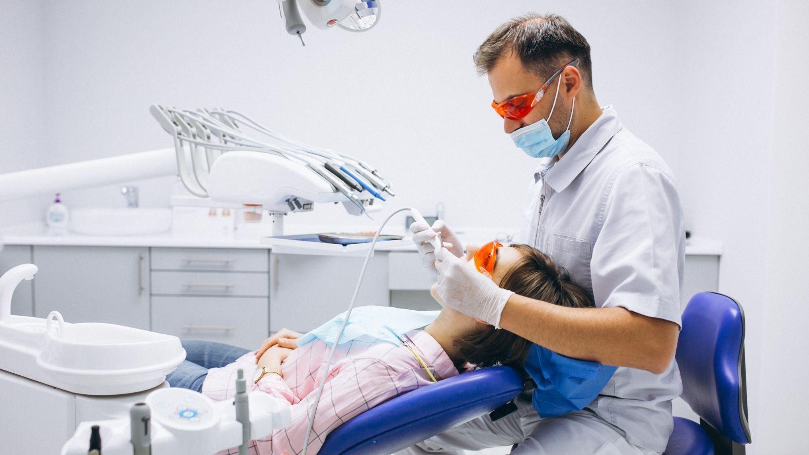
Initial Surgical Response
Right after the implant goes in, bleeding and a blood clot form at the site—this happens within minutes. The clot keeps things stable and releases signaling molecules (cytokines and growth factors) to attract inflammatory and repair cells.
Inflammatory cells like neutrophils and macrophages show up in the first 48–72 hours to clean out bacteria and debris. Macrophages then switch gears and start supporting healing by releasing factors that activate repair cells.
You’ll probably notice some swelling and mild discomfort—honestly, that’s just a sign your body’s immune system is on the job. It’s really important to keep infection under control and avoid disturbing the clot so things don’t go sideways early on.
Cellular Regeneration
Within a few days, mesenchymal stem cells (MSCs) and osteoprogenitor cells move to the implant surface, guided by growth factors. These cells stick to the implant and start turning into osteoblasts, especially if the area stays stable.
At the same time, new blood vessels start forming to bring in oxygen, nutrients, and more cells. If blood supply is weak, bone forms slowly and you risk fibrous tissue sneaking in instead of bone.
Your dentist will probably tell you not to load the implant yet—too much movement can mess up cell attachment. Good oral hygiene, infection control, and a stable environment all help this phase along.
Formation of New Bone
Osteoblasts get to work, laying down osteoid (the unmineralized bone matrix) on and around the implant threads within 1–3 weeks. That osteoid mineralizes quickly, forming woven bone—it’s not super strong, but it fills gaps and gives early contact with the implant.
Woven bone grows in from your own bone and along the implant, slowly increasing the bone-to-implant contact. Honestly, this part takes months—bone volume and density around the implant keep building as things remodel.
Clinicians track this with biochemical markers and x-rays, checking stability before moving on to the next steps. If you’ve had grafts or sinus lifts, this new bone phase might take longer, and your dentist will want to keep a closer eye on things.
Stabilization of the Implant
Over the next several weeks or months, that early woven bone remodels into lamellar (mature) bone with organized collagen and more mineral density. Osteoclasts chew up immature bone while osteoblasts lay down new, stronger bone that lines up with the forces in your mouth.
This remodeling is what shifts the implant from just being held in by a tight fit to being biologically locked in by bone. You might notice the implant feels firmer, and your dentist will likely check stability with torque tests or tapping.
Keeping bone volume up, avoiding too much biting force, and staying clear of infection all matter here. Stick to your post-op instructions and don’t skip those follow-up visits—your implant needs time and care to really lock in.
Key Biological Factors
Osseointegration relies on coordinated cell activity, a good blood supply, and tightly managed inflammation. Each factor shapes how bone forms, remodels, and secures the implant.
Role of Osteoblasts and Osteoclasts
Osteoblasts are your main builders—they lay down new bone matrix right onto the implant. These cells make collagen and mineralize the matrix, creating the first connection that holds the implant steady.
Osteoclasts handle cleanup, removing damaged or extra bone. Their work shapes the bone around the implant, letting it remodel and fit better over time. But if osteoclasts go overboard, they can weaken early stability.
It’s all about balance. Growth factors (like BMPs) and the implant’s surface help osteoblasts do their thing, while RANK/RANKL/OPG signaling keeps osteoclasts in check. Think of osteoblasts as builders and osteoclasts as sculptors—both are necessary, but they have to work together.
Importance of Blood Supply
You need a dense, healthy network of tiny blood vessels at the implant site. Angiogenesis (new vessel growth) brings in oxygen, nutrients, and the cells that actually build the bone.
If blood flow is weak—maybe from poor site prep, disease, or smoking—healing slows down and you risk fibrous tissue forming instead of bone. Good surgical technique that preserves blood vessels and avoids overheating the bone helps keep microcirculation strong.
The research is pretty clear: faster blood vessel growth means quicker bone formation and better implant stability. Things like VEGF levels, oxygen, and even the way the implant fits all influence how well new vessels form.
Inflammatory Mediators
Right after the implant goes in, you get an acute inflammatory phase—that’s your body’s way of setting the stage for healing. Neutrophils and macrophages clear debris and release cytokines (IL-1, IL-6, TNF-α) that draw in stem cells and kick off bone formation.
Macrophages can act as either defenders (M1-type) or healers (M2-type). You want that shift to M2 for tissue repair and bone growth. If inflammation drags on or cytokines stay high, osteoblasts struggle and you risk fibrous tissue instead of bone.
Controlled inflammation also releases growth factors (PDGF, TGF-β) that help with blood vessel growth and osteoblast activity. You can help this phase by keeping things clean, minimizing surgical trauma, and managing any systemic inflammation.
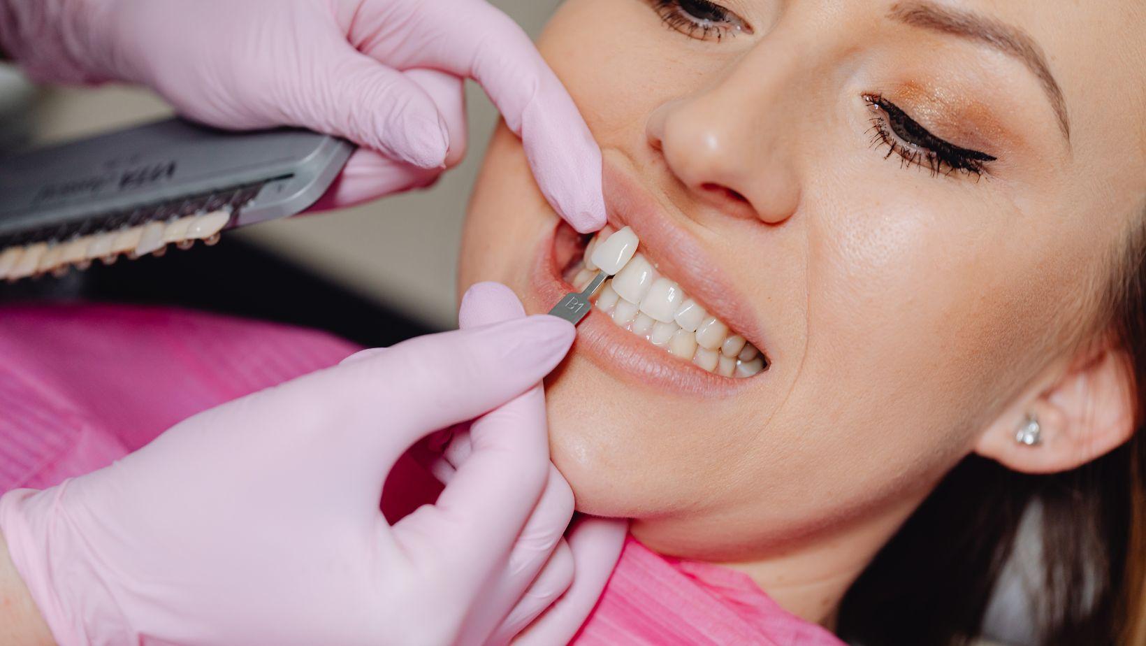
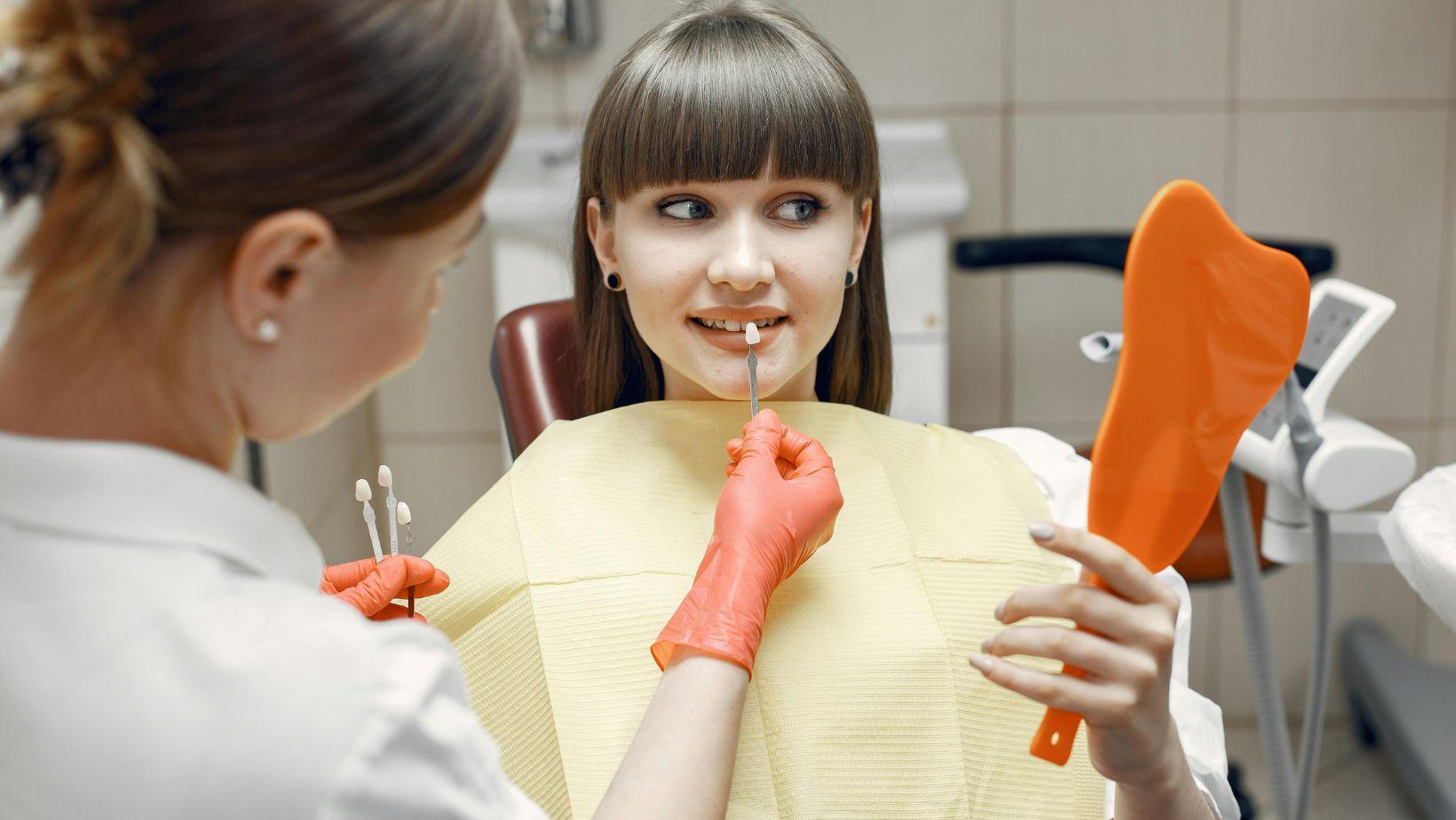
Influence of Implant Materials and Surface Design
The type of implant alloy, surface chemistry, and texture all change how bone cells stick, how fast bone forms, and how stable the implant feels under pressure. Surface roughness, coatings, and the base material each play a role in cell recruitment, protein sticking, corrosion resistance, and mechanical grip.
Surface Texture and Coating
Surface roughness, from big to tiny scales, affects how proteins and cells attach to the implant. Moderate micro-roughness (1–10 µm) helps osteoblasts stick and lay down matrix by boosting surface area and grip. Nanoscale features tweak how proteins land and signal cells to start differentiating.
You’ll see two common coatings: bioactive (like hydroxyapatite or calcium phosphates) and antibacterial or hydrophilic treatments. Bioactive coatings can speed up bone growth by mimicking bone minerals. Hydrophilic or nanostructured surfaces improve wettability, which helps proteins and cells settle in early.
Coating thickness, how crystalline it is, and how well it sticks matter for the long haul. If a coating peels off, it can cause inflammation; on the flip side, well-designed nanoscale roughness improves early integration without sacrificing strength.
Material Biocompatibility
Titanium and its alloys are still the go-to choice—they resist corrosion, are strong yet light, and form a stable oxide layer. That oxide layer protects the surface and limits ion release, which keeps local tissue reactions down.
Other options include zirconia ceramics and some surface-modified polymers. Zirconia collects less plaque and looks better, but it behaves differently under stress and bone response isn’t quite the same as titanium. Polymers are mostly experimental and usually need special surface tweaks to bond with bone.
You’ll want to look at ion release, immune response, and how well the material matches bone’s mechanical properties. Materials that keep inflammation low and have a similar stiffness to bone help avoid stress shielding and support long-term bone health.
Impact on Bone Integration
Surface chemistry and texture guide the whole process—from the first proteins that stick, to cell arrival, bone matrix production, and mineralization. Early events (within minutes to days) depend on how wettable the surface is and what proteins stick; later (weeks to months), it’s all about osteoblast activity and mechanical grip.
Design choices really affect results. Roughened titanium surfaces can cut down the time before you can use the implant, thanks to better early stability. Bioactive coatings can boost bone-to-implant contact in the first months, but only if they stay put. Hydrophilic surfaces help reduce early movement by making clots and cells stick better.
When choosing or designing an implant, you’ll need to balance immediate stability with long-term integration. Your pick should fit the patient’s biology, how soon you want to load the implant, and any risk factors like weak bone or infection.
Factors Affecting Success Rates
Several clinical and biological factors shape how well an implant bonds with bone. Paying attention to systemic health, local bone quality, and surgical details can lower the risk of problems.
Patient Health and Bone Quality
Your overall health plays a big role in osseointegration. Conditions like uncontrolled diabetes, osteoporosis, or smoking can mess with blood flow and bone turnover, slowing healing and raising the chance of failure. Managing things like blood sugar, quitting smoking, and checking meds all help.
The quality of your local bone matters just as much. Dense cortical bone in the front lower jaw usually gives better initial stability than the softer bone in the back upper jaw. If bone is thin or soft, options include grafting, sinus lifts, or using wider or longer implants to get more contact.
Some meds and past radiation can change how bone remodels. Bisphosphonates, antiresorptives, and recent head-and-neck radiation need careful planning—always share your full medical history before starting an implant journey.
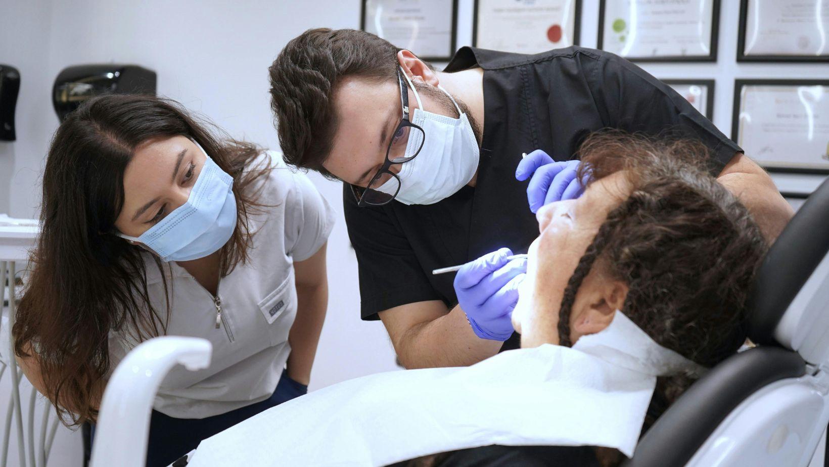
Surgical Technique Considerations
Your surgical approach really shapes both initial stability and how the bone heals. If you use an atraumatic flap design and prep the osteotomy accurately, you’ll help keep the bone healthy.
Avoid overheating during drilling—seriously, it can wreck bone viability and lead to necrosis. I always stick with calibrated drills and plenty of irrigation.
Primary stability is huge, especially if you’re thinking about immediate or early loading. Make sure you hit a torque and insertion value that fits your loading plan.
If torque feels low, it’s better to stage the restoration and let healing take its time. Thread design, implant length, and diameter all play into how well the implant grips the bone.
Sterile technique matters a lot here. I keep infection risks down with solid infection control, and for high-risk patients, I’ll add perioperative antibiotics or chlorhexidine rinses.
Prosthetic planning is the last piece I really focus on. Distributing occlusal loads and designing temporary restorations that don’t stress the healing site can make a big difference.